Master Cardiac Electrophysiology & Action Potentials in 15 Minutes
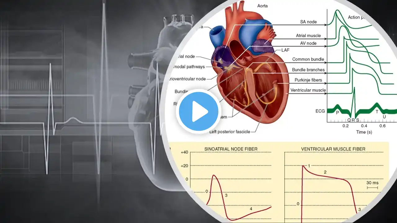
🩺 USMLE Aspirants & Medical Students: Master Cardiac Electrophysiology & Action Potentials in Less Than 15 Minutes! ❤️⚡ This high-yield video breaks down cardiac action potentials, ion channel dynamics & conduction pathways, with insights from Guyton and Hall’s Medical Physiology & Ganong’s Review of Medical Physiology—perfect for USMLE prep, residency training, & clinical mastery! 🔬 Cardiac Action Potentials: The Electrical Blueprint of the Heart Cardiac action potentials regulate heart contractions through precise ion fluxes, ensuring synchronised electrical conduction. ⚡ Pacemaker vs. Contractile Myocytes: Key Differences Pacemaker Cells (SA Node) 🕒 – Spontaneous depolarisation via HCN channels (funny current) Contractile Myocytes 💪 – Require external stimulation to trigger rapid depolarisation 📊 Phases of Cardiac Action Potentials: 1️⃣ Depolarisation (Phase 0) – Fast Na⁺ channels open, causing rapid voltage shift 2️⃣ Early Repolarisation (Phase 1) – K⁺ efflux briefly dominates 3️⃣ Plateau Phase (Phase 2) – L-type Ca²⁺ channels balance K⁺ efflux, crucial for excitation-contraction coupling 4️⃣ Final Repolarisation (Phase 3) – K⁺ channels restore resting potential 5️⃣ Resting Membrane Potential (Phase 4) – Stable at -90 mV in contractile cells, spontaneous depolarisation in pacemaker cells 🔥 Cardiac Conduction System: Keeping the Rhythm SA Node 🕒 – Primary pacemaker, firing ~70 action potentials per minute AV Node 🔄 – Delays impulse for ventricular filling Bundle of His & Purkinje Fibres ⚡ – Ensure rapid ventricular contraction 🧬 Why Cardiac Electrophysiology Matters Understanding action potentials & conduction pathways is essential for diagnosing arrhythmias, mastering clinical physiology, & excelling in USMLE exams!
