How To Measure Femur Length On Ultrasound | FL Measurement Technique | Fetal Biometric Parameter USG
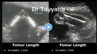
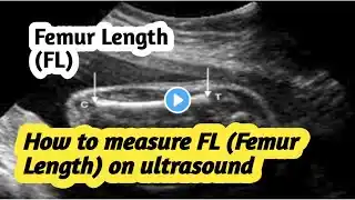
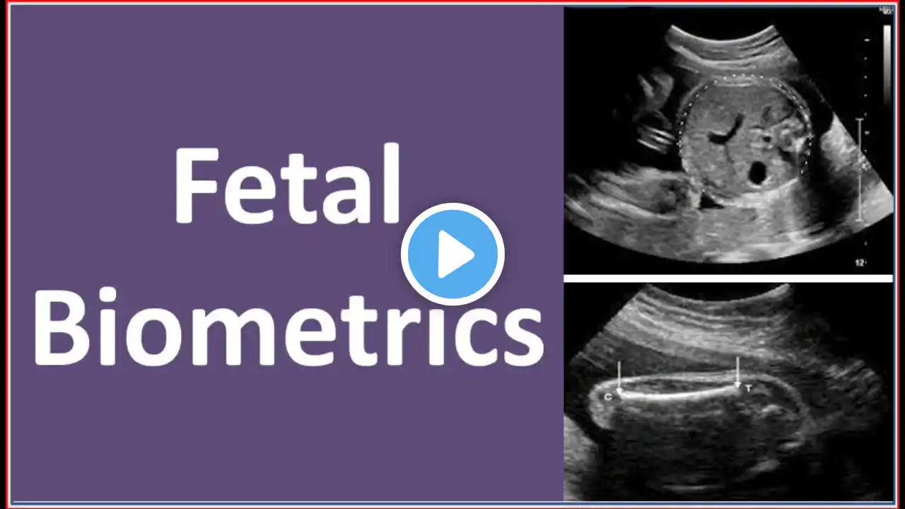
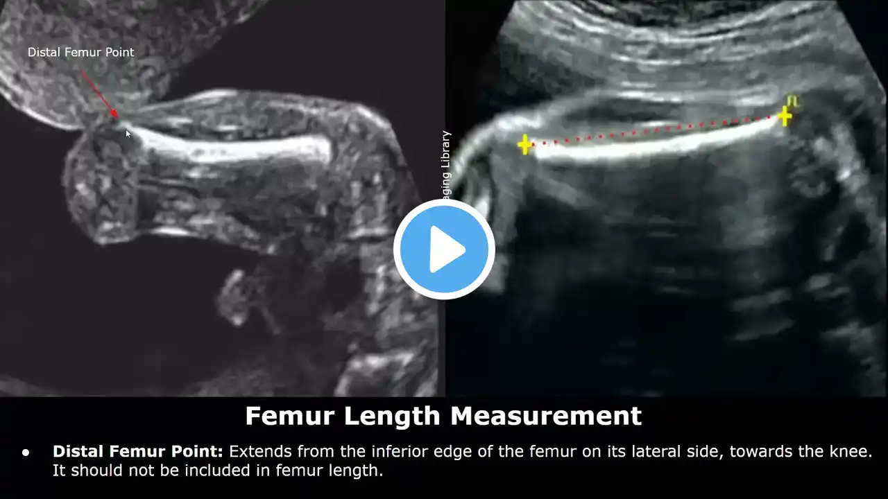
How To Measure Femur Length On Ultrasound | FL Measurement Technique | Fetal Biometric Parameter USG The femur should be imaged as longitudinally as possible, perpendicular to the ultrasound beam. Both ends of the femur (the distal femoral epiphysis and the proximal femoral epiphysis) should be clearly visualized. Avoid excessive pressure with the transducer, which could compress the fetal structures and lead to an underestimation of the measurements. Distal Femur Point: Extends from the inferior edge of the femur on its lateral side, towards the knee. It should not be included in femur length. Position one caliper at the proximal end and the other at the distal end of the femur, where the bone appears brightest and most distinct. It's recommended to take multiple measurements and use the average if there's variability. The measurement should be taken in a straight line, not following the curvature of the bone. Compare the measured FFL to gestational age norms to assess whether the fetus is growing appropriately. A significantly shorter or longer femur compared to the gestational age may suggest potential skeletal dysplasias or other anomalies. However, these measurements can also vary in normal fetuses, so it's important to consider the whole clinical picture, including other biometry measurements and maternal factors.


