Dating Scan In Pregnancy
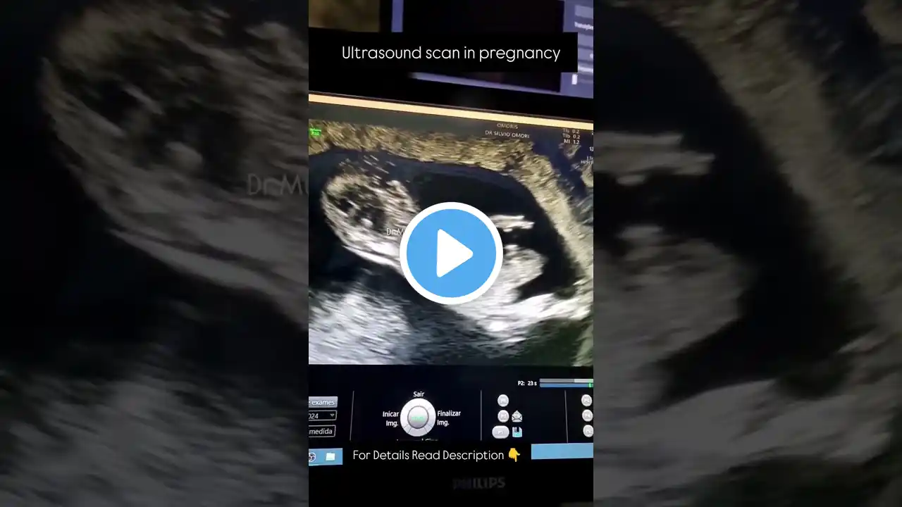
Ultrasound Scan in Pregnancy: Key Information Every Pregnant Woman Should Know 1. What is an Ultrasound Scan in Pregnancy? An ultrasound scan is a non-invasive imaging technique that uses sound waves to create images of the baby inside the womb. It is a standard procedure to monitor the health and development of the fetus during pregnancy. 2. Types of Ultrasound Scans There are several types of ultrasound scans used during pregnancy, including: Transvaginal ultrasound (usually done in early pregnancy) Abdominal ultrasound (common in the second and third trimesters) 3D and 4D ultrasounds (offer more detailed images of the baby) Doppler ultrasound (measures blood flow in the fetus) 3. Purpose of Ultrasound Scans Ultrasounds in pregnancy serve various purposes, including: Confirming pregnancy Determining gestational age Checking for multiple pregnancies (twins, triplets) Monitoring fetal growth and development Evaluating the position of the placenta Assessing amniotic fluid levels Detecting birth defects and abnormalities 4. First Ultrasound: Dating Scan The first ultrasound, often called the dating scan, is typically done between 6 to 9 weeks to confirm the pregnancy, measure the crown-rump length, and determine the due date. 5. Anatomy Scan (20-Week Ultrasound) The anomaly scan, done around 20 weeks, is a detailed ultrasound that examines the baby's organs, bones, and spine for abnormalities. It also checks the placenta, umbilical cord, and amniotic fluid levels. 6. Fetal Heartbeat Detection Ultrasound scans can detect the baby’s heartbeat as early as 6-8 weeks using a transvaginal ultrasound. In later stages, the abdominal ultrasound can monitor the baby’s heartbeat. 7. Ultrasound Safety During Pregnancy Ultrasounds are considered safe for both the mother and baby, as they use sound waves and not radiation. It is a commonly used tool for routine prenatal care. 8. Ultrasound Scans for High-Risk Pregnancies In high-risk pregnancies, ultrasounds may be done more frequently to monitor complications such as low amniotic fluid, placenta previa, or intrauterine growth restriction (IUGR). 9. Detecting Fetal Abnormalities Ultrasound scans can help detect potential fetal abnormalities like spina bifida, Down syndrome, and heart defects, which may require further testing or special care. 10. Monitoring Fetal Position and Movements Ultrasounds can monitor the baby’s position (whether head-down or breech) and track the baby’s movements, which are indicators of the baby’s health and activity. 11. Growth Ultrasound in Late Pregnancy In the third trimester, a growth ultrasound may be done to measure the baby’s size, estimate weight, and ensure that the baby is growing according to expectations. 12. Importance of Ultrasound in Twin Pregnancies For twin pregnancies, regular ultrasounds are important to monitor the growth of both babies, check for twin-to-twin transfusion syndrome (TTTS), and ensure that each baby is developing at a healthy rate. 13. Placenta Health Monitoring Ultrasounds check the placenta’s health, including its position and function. A low-lying placenta (placenta previa) can be identified and monitored through ultrasounds. 14. Amniotic Fluid Check An ultrasound can measure the amniotic fluid index (AFI) to ensure that the baby has enough fluid for optimal development. Too little or too much amniotic fluid can indicate complications. 15. Gender Determination Around 18-20 weeks, an ultrasound can reveal the baby’s gender if desired. However, the accuracy of gender prediction depends on the baby’s position during the scan. 16. Ultrasound and Due Date Estimation While due dates are initially calculated based on the last menstrual period (LMP), early ultrasounds can provide a more accurate estimate of the baby’s arrival date. 17. Doppler Ultrasound for Blood Flow In some cases, a Doppler ultrasound may be used to measure the blood flow in the umbilical cord, baby’s brain, or heart, which can help assess fetal well-being. 18. Preparation for Ultrasound Scans For an abdominal ultrasound, you may be asked to drink water beforehand to have a full bladder, which helps improve image clarity. For a transvaginal ultrasound, no special preparation is needed. 19. Ultrasound Image Interpretation A trained sonographer or obstetrician interprets the ultrasound images. They will guide you through what is visible on the screen and explain the results after the scan. SEO Keywords: Ultrasound scan in pregnancy, types of pregnancy ultrasounds, 20-week pregnancy ultrasound, early pregnancy ultrasound, fetal heartbeat ultrasound, ultrasound for fetal growth, transvaginal ultrasound in pregnancy, ultrasound to detect birth defects, gender determination ultrasound, third trimester ultrasound, 5 weeks ultrasound, 11 weeks ultrasound, Doppler ultrasound pregnancy, safe ultrasounds in pregnancy, fetal movement ultrasound, pregnancy scan